Tests after being diagnosed with uterine (womb) cancer

You may have more tests after your diagnosis to find out:
- How large is the cancer?
- Where exactly is the cancer?
- Has the cancer spread to any other parts of your body?
This is called staging. Staging tests for uterine cancer include:
- Hysteroscopy: A thin, flexible tube with a light at the end (a hysteroscope) is passed through your vagina and into your uterus. This allows your doctor to look inside your uterus and take tissue samples (biopsies). You may be given a local anaesthetic for this test.
- CT scan: This is a type of X-ray that gives a detailed picture of the tissues inside your body.
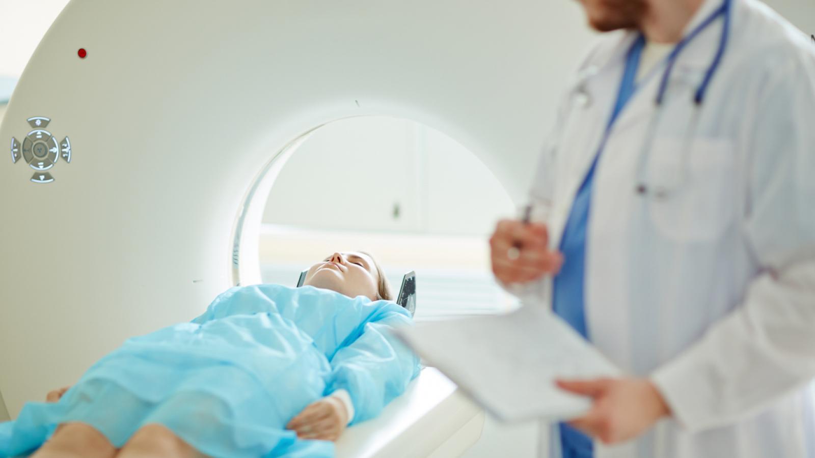
- MRI scan: This is a scan that uses magnetic energy and radio waves to build up a picture of the tissues inside your body. During the scan you will lie inside a tunnel-like machine.
- PET - CT scan: A scan that gives a detailed picture of the tissues inside your body. A low dose of radiotracer (radioactive sugar) is injected into your arm. The scan uses the radiotracer to highlight cancer cells in your body.
- Transvaginal ultrasound: A small metal device called a probe is gently put into your vagina. It uses sound waves to build up a picture of the tissues in your uterus.
Dilatation and curettage (D&C): Here the doctor gently opens your cervix and entrance to the uterus and takes samples of tissue from the inner lining of your uterus. This is done with an instrument shaped like a spoon called a curette. The samples are then sent to the laboratory to be examined. This test is done under general anaesthetic.

Staging is important as it helps your medical team decide on the best treatment for your cancer. Read more about staging tests
For more information
Phone
1800 200 700


