Tests after a lung cancer diagnosis

The tests you have will give your doctors more information about your general health and about the cancer. The tests will help to stage your cancer. This means finding out
- How large is the cancer?
- Where exactly is the cancer?
- Has the cancer spread to any other parts of your body?
Some tests may also be used to see how well your lungs are working and how you are responding to treatment.
Tests you may have include:
Blood tests
Blood tests can help to check your general health. They will be done regularly during your treatment.
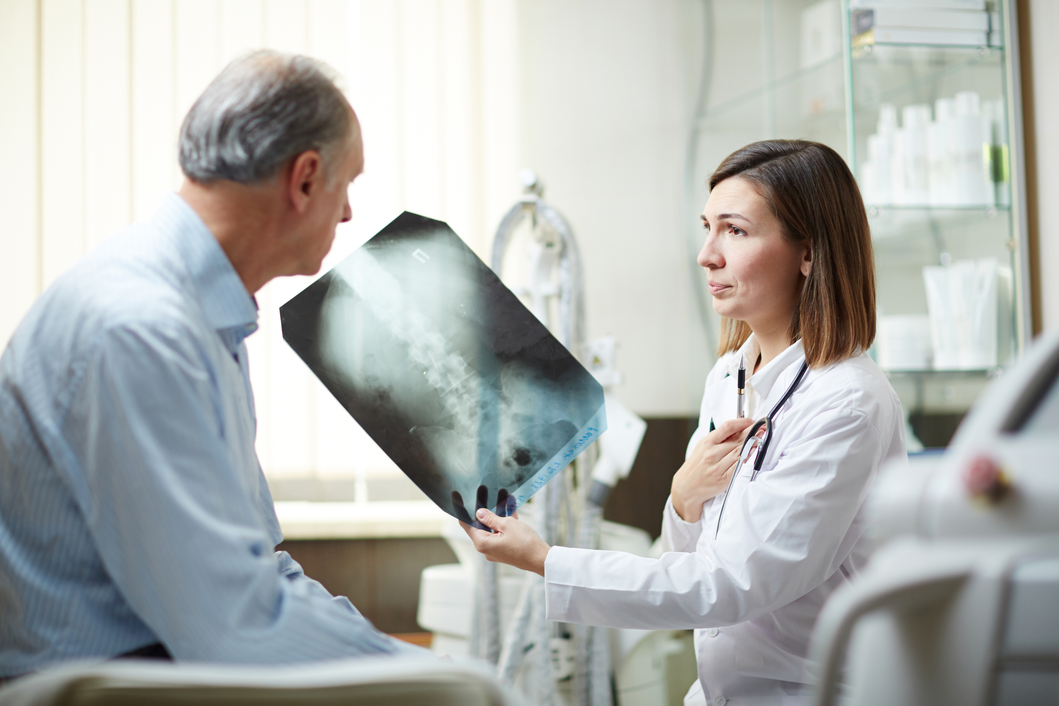
CT scan
A type of X-ray that gives a detailed picture of the tissues inside your body.
Sometimes, a small sample of tissue is taken from your lungs in a test called a CT-guided lung biopsy. The sample is taken using a thin needle and sent to a laboratory for analysis.
As well as a CT scan of your lungs, your doctor may do a CT scan of your abdomen/pelvis and/or brain.
PET - CT scan
A radioactive injection that will show up any cancer spread to other parts of your body on a CT scan picture.
Bronchoscopy
A small tube with a camera on the end is passed into your lungs to take pictures of your lungs. Biopsies (tissue samples) can also be taken during the procedure. Biopsy samples can give information about your type of cancer and how fast it is growing.
Endobronchial ultrasound scan (EBUS)
A type of bronchoscopy that uses an ultrasound probe on the end of the brochoscope to take pictures of the lungs and surrounding lymph nodes. The pictures can show how big the tumour is and whether any nearby lymph nodes are enlarged. Biopsy (tissue) samples from the lung or lymph nodes can be taken by passing a needle through the tube. This is called a transbronchial needle aspiration (TBNA).
Endoscopic ultrasound scan (EUS)
Similar to an EBUS, but the tube with the ultrasound probe goes down your oesophagus (gullet) to give images of the area around the heart and lungs, to show if any of the lymph nodes in the centre of the chest are enlarged.
A fine needle can be passed along the tube to take tissue samples from the lymph nodes.
Mediastinoscopy
Putting a tube with a light and a camera into the centre of your chest area, through a small cut in the front of your lower neck. It can see if there are any abnormal areas.
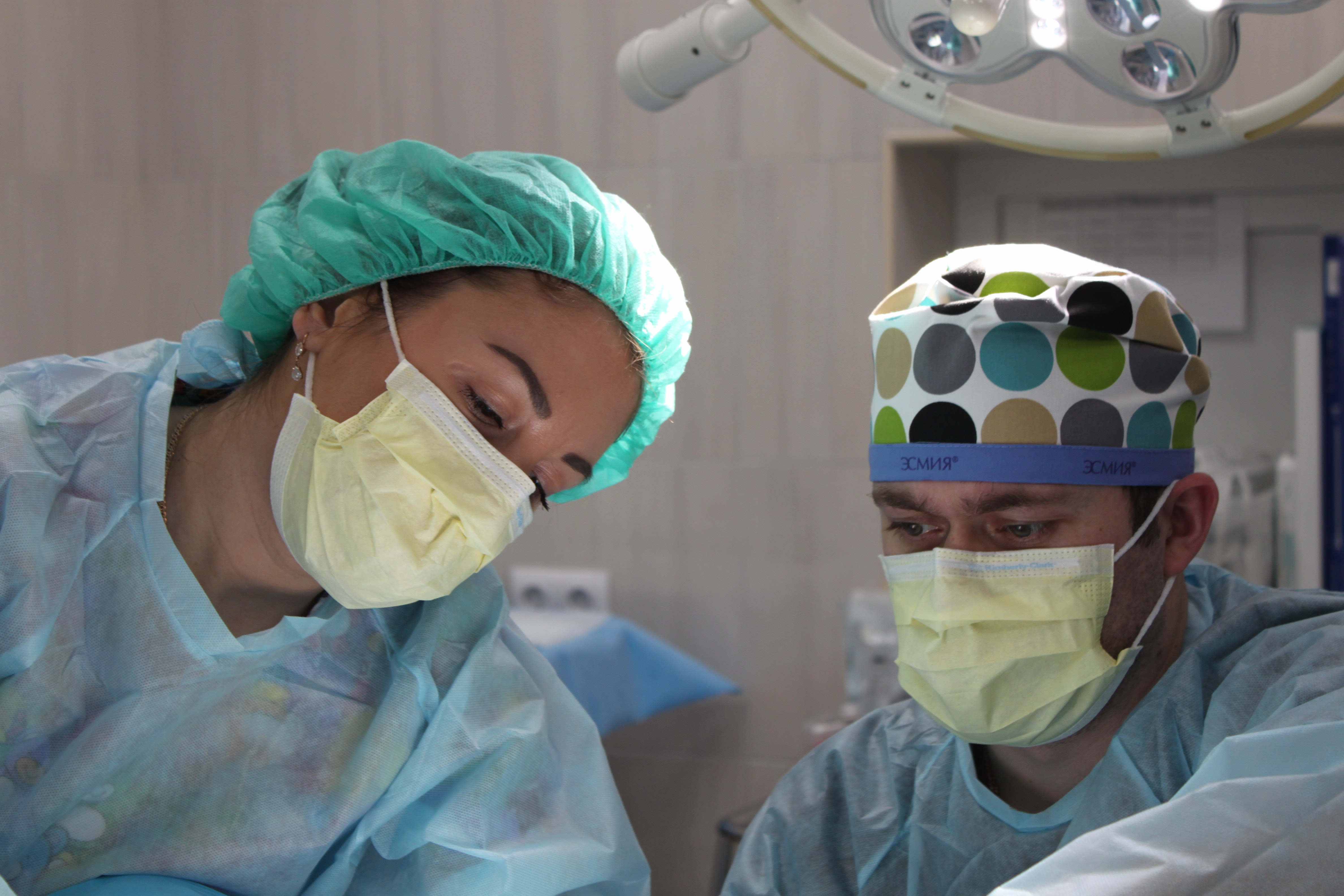
Surgical thoracoscopy
This is a test to look at the membranes that line the lungs (pleura).
A thoracoscope is a long tube with a camera. You will have a general anaesthetic for this operation. Your surgeon will make a cut between two ribs and put the thoracoscope in. This allows them to see if the pleura appear normal.
The surgeon can also take biopsies (tissue samples) of the lining of the inside of the chest and / or the lymph nodes. They can perform a pleurodesis (drain fluid from the lungs), if needed
Bone scan
A radioactive injection that can show areas of abnormal bone on a scan, which may be caused by cancer.
Lung function tests
Your doctor will organise a range of breathing tests to check how well your lungs are working and to see what treatments are possible. The main type of breathing test is a pulmonary function test (PFT), which is where you blow into a mouthpiece on a machine. The test is not painful and takes about 20 minutes. Cardiopulmonary exercise testing (CPET) assesses how your lungs and heart respond to exercise, using an exercise bike
MRI scan
A scan that uses magnetic energy to create a picture of the tissues inside your body. An MRI scan may need to be done to see if the cancer has spread beyond your lung.
MRI scans are not often used for lung cancer unless the cancer is very close to the top of the lung, or other tests suggest an MRI scan is needed.

Staging tests are important as they help your medical team decide on the best treatment for your cancer.
Read more

What are the types of lung cancer?
Asking about your prognosis
How is lung cancer treated?
For more information
Phone
1800 200 700
